Saturday, March 25, 2023

Lupine Publishers| Depression and Anxiety Frequency in Patients Hospitalized on the Guadalajara Regional Military Hospital in the Month of April 2019

 Lupine Publishers| Journal of Neurology and Brain Disorders


Abstract

Observe and Identify patients that presented depression and anxiety using the Hospital Anxiety Diagnosis Scale (HADS), Zung & Conde scale and ASQ 15 scale on Guadalajara Regional Military Hospital during the month of April 01st to April 30th, 2019. Methods: This is a cohort, nonexperimental, observational, prospective and longitudinal study with PubMed and NCBI articles as variables. Findings: Most patients presented anxiety, most patients had a chronic illness, depression was seen mostly in patients older than 50 years old, there was inadequate diet, lack of sleep, and low distress level.

Introduction

Depression and anxiety affect most people around the world, it is characterized by a presence of fear, loss of interest, feelings of guilt or self-esteem that are more commonly associated with sleep disorders, lack of appetite, lack of energy or difficulty concentrating. Depression can become chronic or recurrent and difficult the overall performance on a daily basis, or capacity to live day by day, in its most dangerous form it can lead often to suicide and its lowest form it can be treated with medication and professional psychotherapy [1]. Anxiety is one of the major disorders and its characterized by persistent concern during any activity or routine it is difficult to treat, and it can affect the way a person feels physically [2]. During this investigation we will observe a sample that was taken on the Guadalajara Regional Military Hospital during the month of April a sample of 56 patients presented anxiety and depression according to three scales that were applied.

HADS (hospital anxiety diagnosis scale)

The Hospital Anxiety Diagnosis Scale is an auto applicable questionnaire integrated by 14 items with subscales of seven items one for impared questions and one with pair questions for depression, the authors for this scale are Zigmund and Snaith who proposed this in 1983 and defined the concepts of anxiety and depression the objective of this scale is to identify if the patient has being tensed, concerned or frightened in any way, the 8 items that form the depression subscale are centered around anhedonia with a maximum score that binds from 0 to a 39 score, in which 0-9 score means lack of stress, 10-19 means low stress, 19 to 29 means mild stress and 30 to 39 means anxiety and severe depression.

Zung & conde scale: Its and auto applicable scale consisting of 20 phrases related to depression formed by 10 negative phrases and 10 positive phrases which relate to strong somatic symptoms and 8 cognitive items for each group contemplating the scale with two items referee to mood and other psychotic symptoms [3].

Depression and anxiety: Severe Depression: Its characterized by a combination of symptoms that interfere with capacity to work, sleep, study, eat and enjoy daily basis activities.

Dysthymic disorder: Its characterized by symptoms that is somewhat between 2 years and beyond but less severe, it incapacitates the patient and it prevents him from having a normal life accompanied by a severe depression episode during life [4-6].

Psychotic depression: Occurs during severe depression and its accompanied by some form of psychosis accompanied by delirium and hallucinations.

Seasonal Depression: Its characterized by depression that appears during Winter or times of decreased sunlight.

Bipolar Disorder: Its characterized by maniac depression disorder that its accompanied by cyclic mood swings and depression state, its often seen in patients with cancer, HIV/Aids and Parkinson.

Symptoms

Emotional

Are accompanied by guilt ideas, a severe disease, ideas of sadness never going to heal, loneliness, lack of concentration because patient will eventually die.

Physical

Difficulty eating, or basic needs, weight loss, mood swings.

Negative thoughts

This is mostly seen in older patients, self-stem problems, most cases are seen in patients over 60 years old, or below 45 years old.

Methods

This is a cohort, non-experimental, observational, prospective and longitudinal study in which scholarity was evaluated, cause of hospitalization, age, previous diseases, job and the days patient had been hospitalized.

56 patients both men and women older tan 18 years old were evaluated during this study, a random sample was taken in which every patient has the same possibility of presenting depression or anxiety [6-8]. Patients hospitalized in the women’s hospital room, the men’s hospital room, and the room that consisted of patients that had the rank of major in the Mexican armed forces or above excluding patients that belonged to Intensive care unit, using the Hospital Anxiety Diagnosis Scale, Zung & Conde Scale and ASQ-15 Scale were used during this study (Figure 1-3) [9,10].

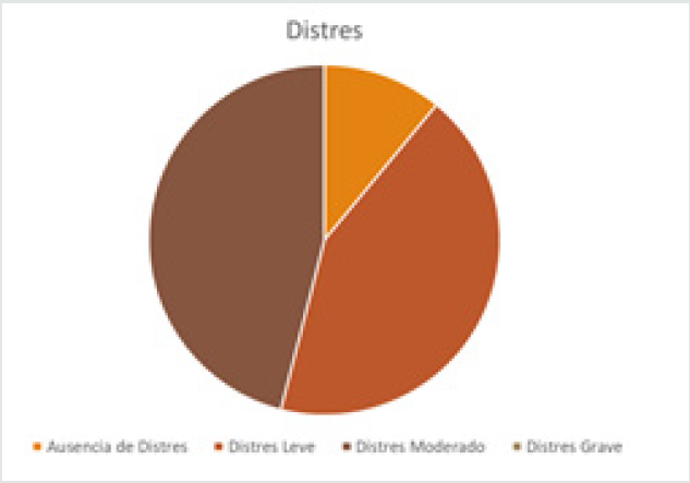
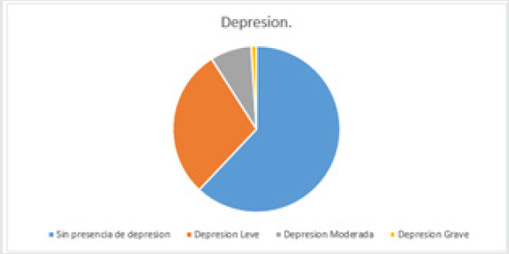
Figure 1: Patients with depression. Fuente. Zung & Conde Depression Scale. 62% of the patients did not present depression 34 patients, 16 patients had low depression 29%, also 8% of the patients had moderate depression which represented 4 patients also 2 of the patients representing 1% presented what could be considered as severe depression.

Lupinepublishers-openaccess-neurology-brain-disorders-journal

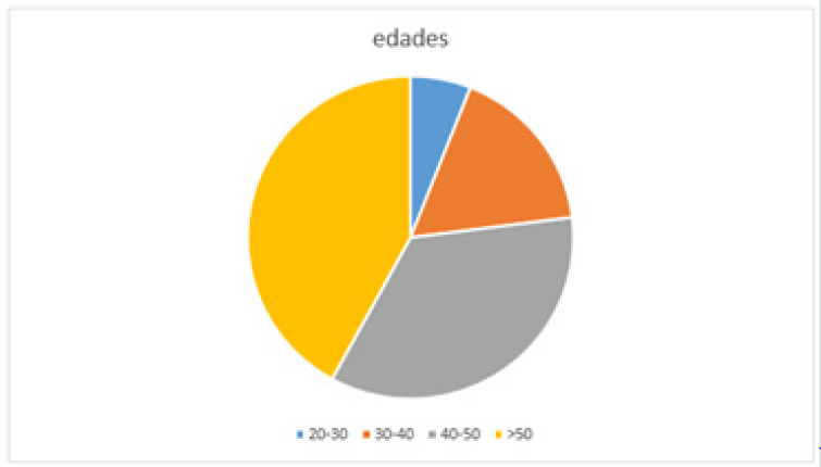
Figure 2: Most common ages seen during study. 42% of the patients presented depression were over 50 years old. 35% of the patients presented depression between 40-50 years of age. 17% of the patients that presented depression had between 30-40 years of age. 6% of the patients had between the ages of 20 to 30 years of age.

Lupinepublishers-openaccess-neurology-brain-disorders-journal

Figure 3: Patients that had some sort of stress evaluated by HADS scale.
a) 43% of the patients had lack of stress.
b) 46% of the patients had low level stress.
c) 11% of the patients had moderate stress.
d) 0% of the patients had severe stress

Lupinepublishers-openaccess-neurology-brain-disorders-journal

Justification

This study was conducted to observe what was the impact of being hospitalized and the relation it had with depression and anxiety in patient, we pretended to find viable date that allowed us to expose the hospital environment and the presence of disease, anxiety and depression (Figure 4) [11,12].

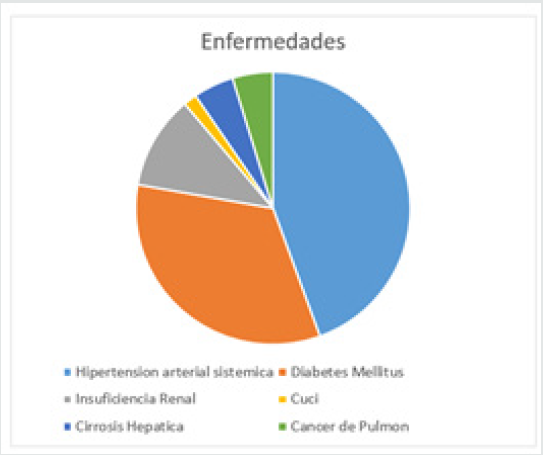
Figure 4: Most common diseases (12 patients didn’t have an illness):
a) Hypertension: 16
b) Diabetes Mellitus:12
c) Renal Insufficiency: 4
d) Ulcerative Colitis:1
e) Hepatic Cirrosis: 3
f) Lung Cancer: 3
g) Cervical Cancer:1
h) Fractures: 4

Lupinepublishers-openaccess-neurology-brain-disorders-journal

Results

Figure 1 Patients with depression. Fuente. Zung & Conde Depression Scale. 62% of the patients did not present depression 34 patients, 16 patients had low depression 29%, also 8% of the patients had moderate depression which represented 4 patients also 2 of the patients representing 1% presented what could be considered as severe depression (Figure 5,6).

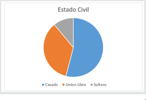
Figure 5: Civil Status:
a) Married: 54%
b) Separated: 35%
c) Single: 11%

Lupinepublishers-openaccess-neurology-brain-disorders-journal

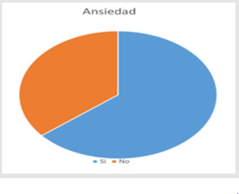
Figure 6: Patients that presented anxiety
a) 64% presented anxiety 35 patients
b) 36% did not present anxiety 21 patients
c) 86% No
d) 14% Yes

Lupinepublishers-openaccess-neurology-brain-disorders-journal

Conclusion

Referred to this subject we understand this two disorders are preventable and the patient if its treated and seeks help with time he can change his daily habits, our suggestions are that a stable lifestyle with a well-balanced diet consisting of fruit, vegetables, meat, daily exercise, stable relationships with family and friends, alongside no work stress, in addition to a good mental stability can lead to a good life and to prevent this type of disorders also to prevent chronic illness which were seen during this study on most patients that indicated feeling anxious or depressed, if they are in this state also to take medication on time and with the help of family members and friend.
a) We observe most patients presented a low depression level
b) Most patients presented anxiety
c) Most patients had a chronic illness
d) Patients over 50 years old presented higher depression levels
e) Most patients were married
Most patients had low level stress level.

Read More about this article: https://lupinepublishers.com/neurology-brain-disorders-journal/fulltext/depression-and-anxiety-frequency-in-patients-hospitalized-on-the-guadalajara-regional-military-hospital-in-the-month-of-april-2019.ID.000171.php

Read more Lupine Publishers Google Scholar Articles: https://scholar.google.com/citations?view_op=view_citation&hl=en&user=X9lN_1AAAAAJ&citation_for_view=X9lN_1AAAAAJ:PcT55Ow6fAIC


Monday, July 18, 2022

Swiss Cheese Pattern a Harbinger of Dementia or an Incidental Finding in an Unusual Case?


Abstract

Dilated Virchow Robin (VR) spaces are pial- line fluid filled structures which surround the walls of small penetrating vessels. In a severe form they develop a swiss cheese pattern or a cribriform pattern in straitum which may predispose to cognitive impairment. We report a patient with change in personality associated with diffuse atrophy, hypometabolism, microbleeds and swiss cheese striatum which is rare.

Read More about this article: https://lupinepublishers.com/neurology-brain-disorders-journal/fulltext/swiss-cheese-pattern-a-harbinger-of-dementia-or-an-incidental-finding-in-an-unusual-case.ID.000170.php

Read more Lupine Publishers Google Scholar Articles: https://scholar.google.com/citations?view_op=view_citation&hl=en&user=X9lN_1AAAAAJ&citation_for_view=X9lN_1AAAAAJ:FKYJxdYMdFIC

Friday, March 25, 2022

Lupine Publishers| Antiaging, Cognition and Anti-Inflammatory Potential of the Biofield Energy Treatment in Vitamin D3 Deficiency Diet (VDD) Induced Sprague Dawley Rats

 Lupine Publishers| Journal of Neurology and Brain Disorders


Abstract

A proprietary formulation was designed that consist of minerals (zinc, magnesium, iron, calcium, selenium, and copper), vitamins (pyridoxine HCl, cyanocobalamin, ascorbic acid, alpha tocopherol, and cholecalciferol), Panax ginsengextract, β-carotene, and cannabidiol isolate. The present study was aimed to evaluate the impact of Consciousness Energy Healing Treatment (the Trivedi Effect®) on a novel test formulation in male Sprague Dawley (SD) rats, fed with vitamin D3 deficiency diet (VDD) for antiaging/cognitive and anti-inflammatory activities. The test formulation was divided into two parts. One part was denoted as the untreated test formulation without any Biofield Energy Treatment, while the other part was defined as the Biofield Energy Treated sample, which received the Biofield Energy Healing Treatment by renowned Biofield Energy Healer, Mr. Mahendra Kumar Trivedi. The level of Klotho protein (anti-aging biomarker) in cerebro-spinal fluids (CSF) was significantly increased by 44.2%, 92.0%, 44.2%, and 43.1% in the Biofield Energy Treatment per se to animals from day -15 (G6), Biofield Energy Treated test formulation from day -15 (G7), Biofield Energy Treatment per seplus Biofield Energy Treated test formulation from day -15 (G8), and Biofield Energy Treatment per seanimals plus untreated test formulation (G9) groups, respectively as compared to the disease control group (G2). The level of β-endorphin in CSF (cognition, pain and inflammation biomarker) was significantly increased by 418.4%, 1155.7% (p≤0.01), 890.4% (p≤0.01), 351%, and 566.7% in the G5, G6, G7, G8, and G9 groups, respectively as compared to the G4. Moreover, serotonin level in CSF was increased by 94.8%, and 63.4% in the G6 and G9 groups, respectively as compared to the G4. The level of 1, 25 (OH)2D3 in CSF was significantly increased by 61.8%, 33.4%, 61.5%, 64.5%, and 30.6% in the G5, G6, G7, G8, and G9 groups, respectively as compared to the VDD induced group (G2). Further, the level of c-reactive protein (CRP, inflammation biomarker) in serum was reduced by 21.2%, 23.1%, 19.8%, 22.4%, and 23.1% in the G5, G6, G7, G8, and G9 groups, respectively as compared to the G2 group. Altogether, results suggested that the Biofield Treated test formulation and Biofield Energy Treatment per se significantly increased antiaging, cognitive, and anti-inflammatory biomarkers that could be helpful in various aging/psychiatric or inflammatory disorders. Thus, the results showed a significant slowdown of disease progression and all other disease-related complications/symptoms in the preventive Biofield Energy Treatment group per seand the Biofield Energy Treated Test formulation groups (viz. G6, G7, G8, and G9) as compared to the disease control group.

Keywords: Biofield Treatment;antiaging; The Trivedi Effect®;klotho;β-endorphin;serotonin; Vitamin D3 deficiency diet;calcitriol

Introduction

Deficiency of vitamin D3 is directly linked to various health problems like osteoporosis, cognitive decline, cardiovascular disease, depression, diabetes, hypertension, and cancer [1,2]. Vitamin D is very essential for bone health in adults and children. Its sufficient concentration prevents osteomalacia, muscle weakness, and protect fractures. The processes by which intake of vitamin D3 like synthesis through skin via UV-rays and absorption from foods become less efficient with age [3]. Hence, hypovitaminosis of vitamin D3 is more prevalence worldwide [4]. Based on this situation authors constructed the current research work to evaluate the impact of Consciousness Energy Healing Treatment on aging after induction of Vitamin D3Deficiency Diet (VDD) in Sprague Dawley rats. The newly formulated test formulation, which is a combination of multiple minerals (iron, copper, zinc, magnesium, calcium, and selenium), vitamins (ascorbic acid, cholecalciferol, pyridoxine HCl, alpha tocopherol, and cyanocobalamin), panax ginseng extract, and cannabidiol isolate. Each component of this test formulation commonly used as nutraceutical supplement [5-8]. Biofield Therapy (or Healing Modalities) is one of the approach of Complementary and Alternative Medicine (CAM) therapies now considering as the first-line model of treatment against several disorders. Based on the obtained data from National Health Interview Survey (NHIS) 2012, reported that most of the Americans used the dietary supplement as complementary health approaches than conventional medicine therapy. Besides, The National Center of Complementary and Integrative Health (NCCIH) has recognized and accepted Biofield Energy Healing as a CAM health care approach in addition to other therapies, medicines and practices such as Tai Chi, Qi Gong, Ayurvedic medicine, Rolfing structural integration, deep breathing, yoga, natural products, chiropractic/osteopathic manipulation, massage, meditation, relaxation techniques, aromatherapy, acupuncture, progressive relaxation, hypnotherapy, healing touch, mindfulness, special diets, naturopathy, homeopathy, guided imagery, acupressure, traditional Chinese herbs and medicines, pilates, movement therapy, Reiki, essential oils, cranial sacral therapy and applied prayer.

Human Biofield Energy has subtle energy that can work effectively [9]. CAM therapies have been practiced worldwide with reported clinical benefits in different health disease profiles [10]. This energy can be harnessed and transmitted by individuals into living and non-living things via the process of Biofield Energy Healing. Biofield Energy Treatment (the Trivedi Effect®) has been published in numerous peer-reviewed science journals with significant outcomes in many scientific fields such as cancer research [11, 12], microbiology and biotechnology [13-15], pharmaceutical science [16-19], agricultural science [20-22], materials science [23-25], dietary supplement [26,27], skin health [28,29], human health and wellness. The planned to evaluate the impact of the Biofield Energy Healing Treatment (the Trivedi Effect®) on the test formulation for antioxidant action concerning lipid peroxidation, antioxidant activity using standard assays.

Materials and Methods

Chemicals and reagents

Calcitriol, pyridoxine hydrochloride (vitamin B6), beta carotene (retinol, Provit A), zinc chloride, and magnesium (II) gluconate were purchased from TCI, Japan. Copper chloride, calcium chloride, cyanocobalamin (vitamin B12), cholecalciferol (vitamin D3), sodium carboxymethyl cellulose (Na-CMC), vitamin E (Alpha-Tocopherol), and iron (II) sulfate were procured from Sigma-Aldrich, USA. Sodium selenate and ascorbic acid were obtained from Alfa Aesar, India. Panax ginsengextract and cannabidiol isolate were obtained from Panacea Phytoextracts, India and Standard Hemp Company, USA, respectively. Other chemicals used in this experiment were analytical grade procured from India.

Experimental animals

Randomly breed male Sprague Dawley (SD) rats with body weight ranges from 200 to 300 gm were used in this study. The animals were purchased from M/s. Vivo Bio Tech, Hyderabad, India. Animals were randomly divided into nine groups based on their body weights consist of 6 animals of each group. They were kept individually in sterilized polypropylene cages with stainless steel top grill having provision for holding pellet feed and drinking water bottle fitted with stainless steel sipper tube. The animals were maintained as per standard protocol throughout the experiment.

Consciousness energy healing strategies

The test formulation was divided into two parts. One part of each ingredient was considered as the untreated test formulation, where no Biofield Energy Treatment was provided. Another part of each ingredient was received Biofield Energy Treatment by Mr. Mahendra Kumar Trivedi (the Trivedi Effect®) under laboratory conditions for ~3 minutes in the research laboratory, Dabur Research Foundation, New Delhi, India. Besides, three group of animals were also received Biofield Energy Treatment under laboratory conditions for ~3 minutes. The energy transmission was done without touching the samples or animals. Similarly, the control samples were subjected to “sham” healer under the same laboratory conditions for ~3 minutes for comparison purposes. The “sham” healer did not have any knowledge about the Biofield Energy Treatment. After that, the Biofield Energy Treated and untreated test formulations were kept in the similar sealed condition and used as per the study plan. The Biofield Energy Treated animals were also be taken back to experimental room for further proceedings.

Experimental procedure

Seven days after acclimatization, animals were randomized and grouped based on body weight. All the animals except G1 were fed with Vitamin D3 deficient diet (VDD) from day -12 to till the end of the experiment. To induce CYP24A1 expression, to accelerate the catabolism of endogenous vitamin D3, the rats (Group G2 to G6) were receive intraperitoneal injections of 40 ng of 19-nor-1,25- dihydroxyvitamin D2 (Paricalcitol) on days -12, -10, -8, -6, -4, -2, day 1, 3 and 5. Group G1 to G5 animals were dosed with respective formulations from Day 1 to till the end of the experiment. However, Group G6 were not be dosed. Animals (50% of the animals from each group) were kept for overnight fasting on Day 56 (Tentative). However, remaining 50% animals were dosed with respective formulations and were kept for fasting on Day 57 (Tentative) next day animals were bled and serum was separated for the estimation of C-reactive protein (CRP). After bleeding, cerebrospinal fluid (CSF) were collected by standard in-house method using stereotaxic instrument for the estimation of KLOTHO, Beta-Endorphin, Serotonin, and 1, 25 (OH)2 D3 by ELISA method.

Estimation of klotho protein, beta-Endorphin, serotonin and 1, 25 (Oh)2 D3 In cerebrospinal fluids (CSF)

The Klotho protein expression was determined using Rat Klotho ELISA Kit in rat’s CSF in according to the manufacturer’s instructions [30].

Assessment of Serum C-reactive protein (CRP)

Serum C-reactive protein were estimated using standard ELISA assay followed by manufacturer instructions. Serum was collected from all the animals after completion of the experiment was examined for level of CRP. The detailed test procedure of the identification of serum C-reactive protein were performed using manufactured instructions as per individual ELISA kit. The CRP level was tested using CUSABIO, ELISA Assay Kit as per manufacturer instructions.

Statistical analysis

The data were expressed as mean ± Standard Error of Mean (SEM) and subjected to statistical analysis using Sigma Plot (Version 11.0). For multiple comparison One-way analysis of variance (ANOVA) followed by post-hoc analysis by Dunnett’s test and for between two groups comparison Student’s t-test was performed. The p≤0.05 was considered as statistically significant.

Results and Discussion

Estimation of klotho protein in Cerebrospinal fluids (CSF)

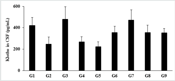
Figure 1: The effect of the test formulation on the level of Klotho protein in cerebrospinal fluids (CSF) in male Sprague Dawley rats. G: Group; G1: Normal control (0.5% CMC); G2: Disease control (VDD: Vitamin D3 deficient diet + 0.5% CMC); G3: Reference item (VDD + Calcitriol); G4: (VDD + untreated test formulation); G5: (VDD + Biofield Energy Treated test formulation); G6: (VDD + Biofield Energy Treatment per se to animals from day -15; G7: (VDD + Biofield Energy Treated test formulation from day -15); G8: (VDD + Biofield Energy Treatment per se plus Biofield Energy Treated test formulation from day -15), and G9: (VDD + Biofield Energy Treatment per se animals plus untreated test formulation). Values are expressed as mean ± SEM, n=6 in each group.
Lupinepublishers-openaccess-neurology-brain-disorders-journal

The impact of the test formulation on the expression of Klotho protein in Cerebrospinal Fluids (CSF) is shown in Figure 1. The level of Klotho protein in the normal control (G1) group was 419.71 ± 75.81 pg/mL and it was decreased by 41.52% in the disease control (G2) group (245.43 ± 69.12 pg/mL) induced by vitamin D3 Deficiency Diet (VDD). The positive control (calcitriol) showed 95.46% increase the level of Klotho protein expression as compared to the G2 group. Further, expression of Klotho protein was significantly increased by 9.31%, 44.24%, 91.97%, 44.24%, and 43.07% in the untreated test formulation (G4), Biofield Energy Treatment per se to animals from day -15 (G6), Biofield Energy Treated test formulation from day -15 (G7), Biofield Energy Treatment per se plus Biofield Energy Treated test formulation from day -15 (G8), and Biofield Energy Treatment per se animals plus untreated test formulation (G9) groups, respectively as compared to the G2 group. Further, the level of Klotho protein was significantly increased by 31.95%, 75.61%, 31.95%, and 30.88% in the G6, G7, G8, and G9 groups, respectively as compared to the untreated test formulation group (G4). Klotho protein acts as an anti-aging biomarker. Klotho gene is recognized as a putative aging-suppressor gene, has a great interest and provides more useful information of the aging process. Data obtained from one experiment in mice reported that the overexpression of the Klotho gene extends the lifespan, and mutations to the klotho gene which shorten the lifespan [31,32].

Assessment of CSF biomarker - β-endorphin

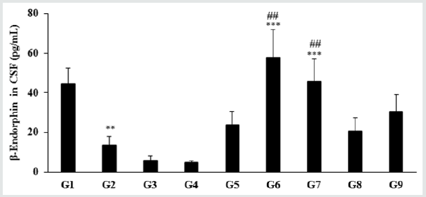
The level of β-endorphin in the normal control group (G1) was 44.48 ± 7.87 pg/mL and it was significantly (p≤0.01) decreased by 69.51% in the disease control (G2) group (13.56 ± 4.36 pg/mL) induced by vitamin D3 Deficiency Diet (VDD). Besides, secretion of β-endorphin was significantly increased by 75.59%, 325.37% (p≤0.001), 235.47% (p≤0.001), 52.80%, and 125.88% in the Biofield Energy Treated test formulation (G4), Biofield Energy Treatment per se to animals from day -15 (G6), Biofield Energy Treated test formulation from day -15 (G7), Biofield Energy Treatment per se plus Biofield Energy Treated test formulation from day -15 (G8), and Biofield Energy Treatment per se animals plus untreated test formulation (G9) groups, respectively as compared to the G2 group. Further, the level of β-endorphin was significantly increased by 418.74%, 1156.64% (p≤0.01), 891.07% (p≤0.01), 351.42%, and 567.32% in the G5, G6, G7, G8, and G9 groups, respectively as compared to the untreated test formulation group (G4). β-endorphin is an endogenous opioid neuropeptide and peptide hormone, considered as cognition, pain and inflammatory biomarker Figure 2. It is produced in certain neurons within the central nervous system and peripheral nervous system to relieve pain when bound to their mu-opioid receptors [33].

Figure 2: The effect of the test formulation on the level of β-endorphin in cerebrospinal fluids (CSF) in male Sprague Dawley rats. G: Group; G1: Normal control (0.5% CMC); G2: Disease control (VDD: Vitamin D3 deficient diet + 0.5% CMC); G3: Reference item (VDD + Calcitriol); G4: (VDD + untreated test formulation); G5: (VDD + Biofield Energy Treated test formulation); G6: (VDD + Biofield Energy Treatment per se to animals from day -15; G7: (VDD + Biofield Energy Treated test formulation from day -15); G8: (VDD + Biofield Energy Treatment per se plus Biofield Energy Treated test formulation from day -15), and G9: (VDD + Biofield Energy Treatment per se animals plus untreated test formulation). Values are expressed as mean ± SEM, n=6 in each group. ***p≤0.001 vs. G2, ##p≤0.01 vs. G4, and **p≤0.01 vs. G1.
Lupinepublishers-openaccess-neurology-brain-disorders-journal

Estimation of 5-hydroxy tryptamine (Serotonin) in CSF

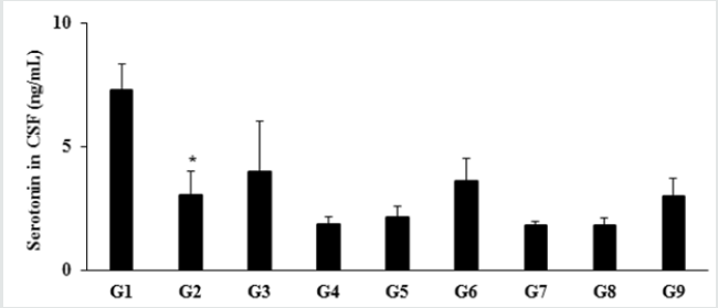
The level of serotonin or 5-hydroxy tryptamine (5-HT) in the normal control group (G1) was 7.29 ± 1.03 ng/mL and it was significantly (p≤0.05) decreased by 58.02% in the disease control (G2) group (3.06 ± 0.93 ng/mL) induced by vitamin D3 Deficiency Diet (VDD). Besides, secretion was increased by 29.41% and 17.32% in the positive control (G3) and Biofield Energy Treatment per se to animals from day -15 (G6) groups, respectively as compared to the G2 group. Further, the level of serotonin was increased by 16.30%, 95.11% and 63.59% in the G5, G6, and G9 groups, respectively as compared to the untreated test formulation group (G4). Serotonin (5-HT) in neuron and neurotransmitter loss leads to aging. The incomplete neurodegenerative processes and serotonergic neurotransmission also leads to aging process [34]. In this experiment, the Biofield Energy Treated test formulation had significantly improve the level of serotonin, which might reduce aging process Figure 3.

Figure 3: Effect of the test formulation on the level of serotonin in cerebrospinal fluids (CSF) in male Sprague Dawley rats. G: Group; G1: Normal control (0.5% CMC); G2: Disease control (VDD: Vitamin D3 deficient diet + 0.5% CMC); G3: Reference item (VDD + Calcitriol); G4: (VDD + untreated test formulation); G5: (VDD + Biofield Energy Treated test formulation); G6: (VDD + Biofield Energy Treatment per se to animals from day -15; G7: (VDD + Biofield Energy Treated test formulation from day -15); G8: (VDD + Biofield Energy Treatment per se plus Biofield Energy Treated test formulation from day -15), and G9: (VDD + Biofield Energy Treatment per se animals plus untreated test formulation). Values are expressed as mean ± SEM, n=6 in each group. *p≤0.05 vs. G1.
Lupinepublishers-openaccess-neurology-brain-disorders-journal

Evaluation of 1, 25 (OH)2 D3 in CSF

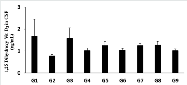
The level of 1, 25 (OH)2 D3 in the normal control group (G1) was 1.67 ± 0.78 ng/mL and it was significantly decreased by 53.89% in the disease control (G2) group (0.77 ± 0.07 ng/mL) induced by vitamin D3 Deficiency Diet (VDD) is shown in Figure 4. The positive control group (G3) had significantly increased the level of 1, 25 (OH)2 D3 by 105.19% compared to the G2 group. Besides, the level of 1, 25 (OH)2 D3 was significantly increased by 31.17%, 62.34%, 33.77%, 62.34%, 64.94%, and 31.17% in the Biofield Energy Treated test formulation (G4), Biofield Energy Treated test formulation (G5), Biofield Energy Treatment per se to animals from day -15 (G6), Biofield Energy Treated test formulation from day -15 (G7), Biofield Energy Treatment per se plus Biofield Energy Treated test formulation from day -15 (G8), and Biofield Energy Treatment per se animals plus untreated test formulation (G9) groups, respectively compared to the G2 group. Further, the level of 1, 25 (OH)2 D3was also significantly increased by 23.76%, 23.76%, and 25.74% in the G5, G7, and G8 groups, respectively.

Figure 4: The effect of the test formulation on the level of 1, 25 (OH)2 D3 in cerebrospinal fluids (CSF) in male Sprague Dawley rats. G: Group; G1: Normal control (0.5% CMC); G2: Disease control (VDD: Vitamin D3 deficient diet + 0.5% CMC); G3: Reference item (VDD + Calcitriol); G4: (VDD + untreated test formulation); G5: (VDD + Biofield Energy Treated test formulation); G6: (VDD + Biofield Energy Treatment per se to animals from day -15; G7: (VDD + Biofield Energy Treated test formulation from day -15); G8: (VDD + Biofield Energy Treatment per se plus Biofield Energy Treated test formulation from day -15), and G9: (VDD + Biofield Energy Treatment per se animals plus untreated test formulation). Values are expressed as mean ± SEM, n=6 in each group.
Lupinepublishers-openaccess-neurology-brain-disorders-journal

Effect of the test formulation on serum CRP level

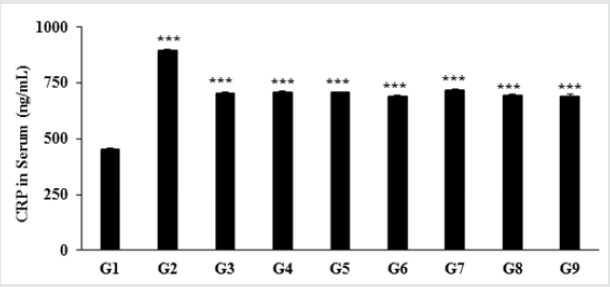
Figure 5: The effect of the Test formulation on change in serum CRP level in vitamin D3 deficiency diet-induced Sprague Dawley rats. G: Group; G1: Normal control (0.5% CMC); G2: Disease control (VDD: Vitamin D3 deficient diet + 0.5% CMC); G3: Reference item (VDD + Calcitriol); G4: (VDD + untreated test formulation); G5: (VDD + Biofield Energy Treated test formulation); G6: (VDD + Biofield Energy Treatment per se to animals from day -15; G7: (VDD + Biofield Energy Treated test formulation from day -15); G8: (VDD + Biofield Energy Treatment per se plus Biofield Energy Treated test formulation from day -15), and G9: (VDD + Biofield Energy Treatment per se animals plus untreated test formulation). Values are expressed as mean ± SEM, n=6 in each group. ***p≤0.001 vs. G1 and G2.
Lupinepublishers-openaccess-neurology-brain-disorders-journal

The effect of the novel test formulation on the level of serum c-reactive protein (CRP) is presented in Figure 1. The serum CRP level in the disease control (vitamin D3 deficiency) group was 895.29 ± 6.02 ng/mL, which was found to be 98.06% higher than that of the normal control (G1) group 452.04 ± 3.80 ng/mL. However, calcitriol group (G3) showed reduced serum CRP level (704.59 ± 6.81 ng/ mL) by 21.30% as compared with the G2 group. The experimental groups such as untreated test formulation to the untreated animals (G4) showed reduced CRP level (708.06 ± 6.37 ng/mL) by 20.91% as compared with the G2 group. Similarly, Biofield Energy Treated test formulation to the untreated animals (G5) reduced the serum CRP level (705.79 ± 4.38 ng/mL) by 21.17% as compared to the G2 group. Biofield Energy Treatment per se to the animals (G6) reduced the CRP level (688.59 ± 6.46 ng/mL) by 23.09% lower as compared to the G2 group. In addition, 15 days pre-treatment of Biofield Energy Treated test formulation (G7) reduced the CRP level (718.14 ± 2.95 ng/mL) by 19.79% as compared to the G2. Another group, 15 days pre-treatment of Biofield Energy Treated test formulation to the Biofield Energy Treated animals (G8) reduced the CRP level (694.41 ± 5.89 ng/mL) by 22.44% as compared to the G2. Similarly, the untreated test formulation to the Biofield Energy Treated animals (G9) reduced the CRP level (688.61 ± 13.29 ng/ mL) by 23.09% as compared to the G2 group. CRP is one of the major inflammatory biomarkers (highly sensitive protein) for inflammatory disorders [35,36]. Thus, Biofield Energy Treatment per se and the test formulation significantly reduced the serum CRP, which significantly improve the inflammatory conditions Figure 5.

In this research plan, four groups were considered as preventive maintenance groups. These groups were G6 (Biofield Energy Treatment per se to animals at -15 days), G7 (Biofield Energy Treated test formulation from day -15), G8 (Biofield Energy Treatment per se to animals along with Biofield Treated test formulation from day -15), and G9 (Biofield treatment per se at -15 days to animals with untreated test formulation). The results showed a significant slowdown of disease progression and all other disease-related symptoms/complications and also reduced the chances of disease susceptibility in these groups. Specifically, group G6 (preventive Biofield Energy Treatment group per se at -15 days) showed the best results as a preventive treatment group compared to the other groups. Based on the overall data, it suggests that the Biofield Energy Healing Therapy was found to be most effective and beneficial to prevent and protect from the occurrence of any type of disease in the rat model. The data indicated that this therapy could act as a preventive maintenance therapy to prevent the occurrence of disease, slow down the disease progression when disease-related complications are present which will ultimately improve the overall health and quality of life.

Conclusion

Results of the study revealed that the level of Klotho protein (anti-aging biomarker) in cerebro-spinal fluids were significantly increased by 44.2%, 92.0%, 44.2%, and 43.1% in the Biofield Energy Treatment per se to animals from day -15 (G6), Biofield Energy Treated test formulation from day -15 (G7), Biofield Energy Treatment per se plus Biofield Energy Treated test formulation from day -15 (G8), and Biofield Energy Treatment per se animals plus untreated test formulation (G9) groups, respectively as compared to the disease control group (G2). Moreover, the level of β-endorphin (cognition, pain and inflammation biomarker) was significantly increased by 418.4%, 1155.7% (p≤0.01), 890.4% (p≤0.01), 351%, and 566.7% in the G5, G6, G7, G8, and G9 groups, respectively as compared to the G4 group. Moreover, serotonin level was increased by 94.8%, and 63.4% in the G6 and G9 groups, respectively as compared to the G4. Further, 1, 25 (OH)2 D3 was significantly increased by 61.8%, 33.4%, 61.5%, 64.5%, and 30.6% in the G5, G6, G7, G8, and G9 groups, respectively as compared to the VDD induced group (G2). The level of c-reactive protein (CRP, inflammation biomarker) was reduced by 21.2%, 23.1%, 19.8%, 22.4%, and 23.1% in the G5, G6, G7, G8, and G9 groups, respectively as compared to the G2 group. The current findings conclude that the Trivedi Effect®-Biofield Energy Healing Treatment has significantly enhanced the antiaging, cognitive, and anti-inflammatory biomarkers level that could be helpful in various aging/psychiatric or inflammatory disorders. which can also be used to improve the overall health. Biofield Energy Healing Treatment (The Trivedi Effect®) per se showed the best results with respect to different beneficial efficacy and biomarker parameters in the preventive maintenance group, G6, as compared to the other preventive maintenance groups (G7, G8, and G9) in the rat model study.

The Biofield Energy Healing Treatment also helped to slow down the disease progression and disease-related complications impacting the overall animals’ health. These data suggested that Biofield Energy Treatment per se and Biofield Energy Treated Test formulation in combination would be the best treatment strategy to prevent and protect from the occurrence of any type of disease. Therefore, the Biofield Energy Healing Treatment (the Trivedi Effect®) per se might be effective in healthy humans when used as a preventive maintenance therapy to sustain good health, to boost overall health, promote healthy aging and increase quality of life. In the presence of disease, the Biofield Energy therapy might reduce the severity of any acute/chronic disease (such as auto-immune related and inflammatory disorders) and / or slow the disease progression. Thus, the Biofield Energy Treated test formulation may act as an effective anti-inflammatory and immunomodulatory product for various autoimmune disorders such as Addison Disease, Systemic Fibromyalgia, Lupus Erythematosus, Hashimoto Thyroiditis, Celiac Disease (gluten-sensitive enteropathy), Multiple Sclerosis, Dermatomyositis, Graves’ Disease, Pernicious Anemia, Aplastic Anemia, Type 1 Diabetes, Myasthenia Gravis, Crohn’s Disease, Vasculitis, Scleroderma, Rheumatoid Arthritis, Psoriasis, Reactive Arthritis, Sjogren Syndrome, Chronic Fatigue Syndrome, Vitiligo, and Alopecia Areata, as well as inflammatory disorders such as Irritable Bowel Syndrome (IBS), Asthma, Ulcerative Colitis, Parkinson’s Disease, Alzheimer’s Disease, Dermatitis, Atherosclerosis, Hepatitis, and Diverticulitis. Further, the Biofield Energy Healing Treated test formulation can also be used in the prevention of immune-mediated tissue damage in cases of organ transplants like kidney transplants, heart transplants, and liver transplants, and in the improvement of overall health and quality of life.
 Read More Lupine Publishers Journal of Neurology and Brain Disorders: https://brain-disorders-lupine-publishers.blogspot.com/

Friday, February 25, 2022

Lupine Publishers| Schizophrenia, Carbonyl Stress and Carnosine

 Lupine Publishers| Journal of Neurology and Brain Disorders


Abstract

Recent research suggests that schizophrenia is associated with the development of an advanced aging phenotype (carbonyl stress) and erythrocytes from schizophrenics also exhibit symptoms of cellular aging (increased levels of glycated proteins and ubiquitinated proteins), possibly due to excessive glycolysis-induced methylglyoxal (MG) generation. The endogenous dipeptide carnosine (beta-alanyl-L-histidine), which can delay cellular aging, suppress glycolysis and inhibit MG-induced protein glycation, also exerts some beneficial effects towards schizophrenia. Carnosine is present in human erythrocytes and the olfactory bulb (olfactory dysfunction is associated with schizophrenia). It is suggested that enhanced erythrocyte and olfactory carnosine levels may be more therapeutic towards schizophrenia, if carnosine was also administered intra-nasally to avoid serum carnosinase activity.


Keywords:Carnosine; glycation; methylglyoxal; erythrocyte; aging; nasal administration

Introduction


Schizophrenia and carbonyl stress

Many studies have indicated a relationship between schizophrenia and dysfunctional energy metabolism [1-3] whilst others indicate that carbonyl stress and generation of advanced glycation end-products (AGEs) accompany schizophrenia [4,5]. Furthermore, a recent study suggests that changes in glycolysis and accelerated cellular aging in glial cells contribute to the condition [6]. The glycolytic intermediates glyceraldehyde-3-phosphate and dihydroxyacetone-phosphate are the most likely sources of AGE formation due to their ability to spontaneously decompose into methylglyoxal (MG). MG is well recognized as a major glycating agent and is thought to be responsible for much macromolecular modifications associated with type-2 diabetes and age-related neurodegenerative conditions [7,8]. However, there is no clear evidence whether suppression of MG generation, via decreased glycolytic activity, has any effect on schizophrenia. The suggestion that schizophrenia seems to be associated with accelerated cellular aging [6] is supported by another recent observation reporting that erythrocytes obtained from schizophrenics contain elevated mounts of ubiquitinated proteins [9]. This might arise from either increased generation of targets for ubiquitination (e.g. aberrant polypeptides or denatured misfolded proteins), or decreased de-ubiquitinating activity, or decreased proteasomal proteolytic activity which would normally complete polypeptide destruction. Interestingly, MG and other agents responsible for carbonyl stress, also induce protein cross-linking which not only renders the target protein less susceptible to proteolytic attack but can also result in inhibition of proteasome activity generally [10]. Thus, it is conceivable that excessive glycolysis can provoke an aging phenotype (AGE accumulation and proteostatic dysfunction) via increased MG generation; such a relationship has been demonstrated in mice fed a high glycemic- index diet [11]. Never-the-less it is necessary to show whether glycation compromises proteostatic in erythrocytes from schizophrenics.


Erythrocytes and schizophrenia

A number of recent papers have revealed that erythrocytes obtained from patients with neurological problems, such as Alzheimer’s Disease (AD) and Parkinson’s Disease (PD), exhibit symptoms typical of aging cells in general. For example, compromised proteolytic activity and MG detoxification were detected in AD erythrocytes [12] and accumulation of aggregated protein occurs in red cells from PD patients [13]. Furthermore, dysfunctional energy metabolism, especially in relation to glycolysis culminating in carbonyl stress, are now regarded as characteristics of both AD and PD [14,15]. Therefore, it is not surprising that evidence of carbonyl stress is also accompanied by enhanced protein glycation [16] and accumulation of ubiquitinated proteins [9] in erythrocytes (and possibly other cells) obtained from schizophrenic individuals [17]. Moreover, one of the glycated proteins from “schizophrenic” red cells has been identified as a selenium-binding protein (SBP1) [18]; dysfunctional selenium metabolism has long been regarded as an important contributor to schizophrenia [19,20]. Selenium plays an important role in Sulphur metabolism required for synthesis of antioxidant enzymes such as glutathione peroxidase [21]. Thus, one is beginning to understand the relationship between AGE generation, carbonyl and oxidative stress and the apparently disparate biochemical attributes to schizophrenia.


Carnosine, carbonyl stress and schizophrenia

That erythrocytes can contain elevated amounts of MG and glycated proteins suggests the possibility that such red cells could become systemic sources of MG and AGEs to the brain and other tissues, following MG-induced eryptosis [22]. Consequently, it is important to consider whether suppression of carbonyl stress, not only in erythrocytes but in astrocytes and glia, could possibly be a therapeutic strategy. The naturally occurring dipeptide carnosine (beta-alanyl-L-histidine) has been shown to suppress glycolysis in cultured cells [23,24], delay replicative senescence [25], stimulate proteolysis of long-lived proteins in late passage cells [26] and inhibit AGE formation [27]. Furthermore, there is one study showing that schizophrenics subjected to dietary supplementation with carnosine exhibited some beneficial effects [28], possibly due to the dipeptide’s pluripotent properties [29]. It is also interesting to note that

a) Olfactory dysfunction is also associated with schizophrenia [30,31] and

b) Carnosine is enriched in the olfactory bulb [32].

Thus, one has to consider whether raising olfactory carnosine levels could also be useful. However, all studies employing dietary carnosine supplementation are subject to the problem of the presence of serum carnosinase activity which would destroy the dipeptide [33]. There is an alternative route however, which is to use an intra-nasal approach. This could involve a nasal spray of a carnosine solution; another approach could involve use of carnosine powder. Indeed “snorting” carnosine could be far more useful than most white powders some people use, be it illegal drugs or “medicinal snuff “of old. In fact, intra-nasal delivery of potential therapeutic agents is currently being explored [34] with respect to neurodegenerative conditions, as proposed many years ago [35].

Carnosine has been detected in human erythrocytes [36] but in lower amounts when obtained from elderly individuals [36]. It is presumed that red cell carnosine is synthesized (from betaalanine and histidine) during erythropoiesis. Consequently, it would be useful to determine whether dietary supplementation with carnosine or beta-alanine raises erythrocyte carnosine levels and whether there are any beneficial effects with respect to the recognized changes in “schizophrenic” erythrocytes. Additionally, it is suggested that any carnosine (dietary or nasally administered) supplementation period should last for at least 120 days to ensure maximal numbers of carnosine-enriched erythrocytes. It has been proposed that excessive and continuous glycolysis in erythrocytes enhances red cell MG levels, and thus also facilitate delivery of erythrocyte MG to the tissues including the brain [22]. Consequently, it will be also important to determine whether such supplementation protocols decrease carbonyl stress and MG levels not only in red cells but the tissues generally including glia [6].

  Read More Lupine Publishers Journal of Neurology and Brain Disorders: https://brain-disorders-lupine-publishers.blogspot.com/

Friday, February 4, 2022

Lupine Publishers| Primary Lateral Sclerosis, Report of a Case and Bibliography Revision

 Lupine Publishers| Journal of Neurology and Brain Disorders

Abstract

Introduction: Primary lateral sclerosis is a rare disease involving the upper motor neuron, producing a bulbospinal spasticity. The course of the disease is insidious and progressive, usually starting with the lower extremities, and later becoming a tetrapyremidal syndrome. As a rare disease, diagnosis in most cases is exclusionary, and the patient should be studied extensively, clinically, including a thorough medical history, laboratorial and with the relevant cabinet studies.

Classic Case: It is male patient who starts his clinical picture about a year ago with weakness in left pelvic limb, subsequently accompanied by pain and paresthesias, manifesting the same symptomatology later in the contralateral leg and upper left limb. Currently is added index and middle toe hypoesthesia, moderate tremor in left arm, with the contrast of the middle toe over the ring of said hand. It has an inability to lift light objects for short periods of time, as well as fatigue in short periods of time when performing daily activities, which greatly limits their daily life.

Conclusion: Motor neurone diseases are divided into two groups, and in the case studied, the upper motor neuron is exclusively affected. As it is a rare disease, with a low incidence, multiple differential diagnoses will be considered before concluding in it, considering it a diagnosis of exclusion. The natural history of the disease will always have a grim outcome, with poor prognosis for life and function, despite the measures taken to change the course of the disease.

Keywords:Motoneuron; upper; spastic; resonance

Introduction

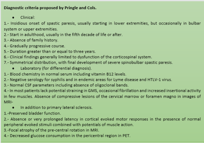
Primary lateral sclerosis is a rare disease involving the upper motoneuron, which is characterized by a progressive bulbospinal spasticity, with selective degeneration of pyramidal neurons located in the precentral convolution [1]. Primary lateral sclerosis makes up approximately 1% to 4% of all patients with motor neuron diseases [2]. The onset of the disease is insidious, with a slow and progressive spastic paralysis, usually starting at the lower extremities, and then becoming a tetrapyremidal syndrome. On average, patients are estimated to have a life prognosis greater than 10 years from the onset of symptoms. Hyperreflexia, moderate weakness, dizziness, lack of coordination may also occur within the clinical picture. In the physical examination you can find spasticity, increased osteotendinous reflexes, and as a predominant sign, stiffness [2,3]. Diagnosis is usually performed clinically, relying on MAGNETIC resonance imaging, among other studies, to rule out other pathological entities, showing in this study a marked atrophy in the primary motor cortex [3]. In some studies, diffusion magnetic resonance imaging has been used, showing a functional increase in cerebral axonal activity, especially the brain-cerebellar, which could explain an adaptive process through functional neuroplasticity, however, the prognosis of the disease remains unfavorable and irreversible despite these changes [4]. Although there are some diagnostic criteria for the disease, as it is a rare entity, not all inclusions for it are always met, so it is usually a diagnosis of exclusion [5,6] (Figure 1).

Figure 1: Diagnostic criteria proposed by Pringle and Cols.

Lupinepublishers-openaccess-neurology-brain-disorders-journal

Clinical Case

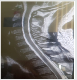
They are a 42-year-old male patient, who started his clinical picture about a year ago, when when he is parading, I notice some weakness in left pelvic limb, progressing over the course of the days to diffuse pain and local paresthesias, and subsequently having the same problem in the contralateral leg and then to upper left limb throughout, progressively to date. Currently, adding to the above, it has hypoesthesia of index and middle toe, stiffness in all the arcs of the movement of the affected limbs, moderate tremor in the left arm, with the oversetting of the middle toe over the ring of said hand. Symptomatology is exacerbated in the mornings. It has an inability to lift light objects for short periods of time, as well as fatigue in short periods of time when performing daily activities, which greatly limits their daily life. It denies important here family backgrounds, as well as surgeries, drug addictions, or known diseases (Figure 2).

Figure 2: Cervical spinal MRI where you see a normal morphology without compromise spinal cord.

Lupinepublishers-openaccess-neurology-brain-disorders-journal

Discussion

Primary lateral sclerosis is a rare disease affecting the upper motor motoneuron of the primary motor area (pre-central circumvolution),characterized by having an insidious onset in people with no known risk factors, with a clinical picture in which a spastic paresis of lower limbs of onset predominates that can be generalized until tetra pares progressively increasing over the years and which is usually a long-evolving disease. Diseases of the motor neuron are divided into those that affect the upper and lower, in this case we focus on the first case, which are located inside the cerebral cortex and send axons that form the pyramidal pathway, to later defuse and finally connect to the spinal cord. The manifestations secondary to the lesion of the upper motoneuron are as follows: spastic paralysis, amyotrophy (by disuse), absence of tracing, exalted myotatic reflexes and extensive plantar response.

As it is a disease with a low incidence and few reported cases, it should never be considered as a diagnosis of first instance, having to rule out other differential diagnoses, so over time various diagnostic criteria have been defined to realize the disease, however, as well mentioned, it is a rare disease, so no definitive consensus has been created for the diagnosis and scrutiny of the disease, however, we can lead our diagnostic approach means of any of these, such as the criteria of pringle and cabbage, through a well-established clinic, conducting a thorough clinical history, laboratory studies, ranging from general analyses such as hematic biometry, blood chemistry, functional tests, to cytological examinations of cerebrospinal fluid, quantification of vitamin B12 in serum, trepamic tests, as well as cabinet studies such as electromyography and MRI, where the absence of other pathological entities is verified and in the latter study a marked cortical atrophy of the pre-central turn. Despite the above, it should be noted that the disease may not follow a specific pattern within its natural history, so it may vary the presentation and sequence of them.

Conclusion

Motor neuron diseases are divided into two groups, and in the case studied, the upper motoneuron is exclusively affected, differing from each other by the form of presentation and clinical manifestations. As it is a rare disease, with a low incidence, multiple differential diagnoses will be considered before concluding in it, considering a diagnosis of exclusion, and thus, a thorough medical history should be made with a proper examination conducting laboratory and cabinet studies relevant to this situation. Although it is a long-term disease with a longer life expectancy than amyotrophic lateral sclerosis, the natural history of the disease will always have a grim outcome, with poor prognosis for life and function, taking into account there is no cure for the disease other than supportive treatment.

 Read More Lupine Publishers Journal of Neurology and Brain Disorders: https://brain-disorders-lupine-publishers.blogspot.com/


Friday, December 10, 2021

Lupine Publishers| Objective Structured Examinations as Supplemental Equipment for Amending Emotional Intelligence: A Pilot Survey

 Lupine Publishers| Journal of Neurology and Brain Disorders

Abstract

Background: The scientific study of emotional intelligence (EI) in organizations has gained considerable research activity over recent years because it is being concerned with awareness and management of one’s own feelings and emotions in daily living activities. The objective of the present study was to investigate the relationship between EI of a group of psychiatric residents and their academic achievement to see that whether proper training and evaluation by new educative instruments can recompense any degree of shortage in EI.

Methods: Consistent with a cross-sectional survey design, 31 psychiatric residents had been requested to answer to The Schutte Self Report Emotional Intelligence Test (SSEIT), in June 2014, for examining the situation with respect to objective structured examinations, like mini-Clinical Examination Exercise (mini-CEX), Objective Structured Clinical Examination (OSCE), and chartstimulated recall (CSR) scores, which had been taken in the earlier 6 months. SSEIT score of 90 had been taken as demarcating point for dividing the sample population into two parallel groups, including the first group with SSEIT score lower than 90 and second group with SSEIT score equal to or more than 90.

Results: The response rate was 93.54%. In line with the results, there was no meaningful relationship between the aforesaid first group and second group as regards the relationship between SSEIT’s score and the mean total score of Mini-CEX, OSCE, and CSR, which had been examined in the preceding 6 months.

Conclusion: The current study demonstrates that EI does not seem to be a fixed problem in psychiatric residents, and enough exercise along with improvement of necessary interrogating or clinical skills may improve or compensate for unsatisfactory EI.

Keywords:Emotional Intelligence; Objectives Structural Examinations; Psychiatric Residents


Introduction

The scientific study of Emotional Intelligence (EI) in organizations has gained considerable research activity over recent years [1]. Simultaneously researchers have investigated and raised concerns about the appropriate way to measure EI in various studies [2]. Although EI has been the subject of much attention at both popular and academic level, only now are answers provided to some of the fundamental questions posed about the construct [3]. Dulewicz, Higgs and Slaski confirm that in literature there appears to be some debate about what constitutes the domain of EI, about terminology used to describe the construct and about methods used to measure it [4]. One method that has been used widely in research to measure EI is the Schutte Emotional Intelligence Scale (SEIS) [5]. Dulewicz et al. state that EI is not a new concept [4]. Mayer, Salovey and Caruso [6] define the concept of EI as the capacity to reason about emotions, and of emotions to enhance thinking. EI includes the abilities to accurately perceive emotions, to access and generate emotions in order to assist thoughts, to understand emotions and emotional knowledge, and to reflectively regulate emotions in order to promote emotional and intellectual growth [6]. Dulewicz and Higgs [4] define EI as being concerned with being aware of and managing one’s own feelings and emotions; being sensitive to and influencing others; sustaining one’s motivation; and balancing one’s motivation and drive with intuitive, conscientious and ethical behavior. It is apparent that from this theoretical perspective EI refers specifically to the co-operative combination of intelligence and emotion [7]. EI emphasizes the importance of self-awareness and understanding, redressing a perceived imbalance between intellect and emotion in the life of the collective Western mind [7]. Zeidner et al. further state that EI also connects with several cutting-edge areas of psychological science, including the neuroscience of emotion, self-regulation theory, studies of meta-cognition, and the search for human cognitive abilities beyond ‘traditional’ academic intelligence. Given the core proposition that it is a combination of IQ and EI that determines life success, a question arises as to whether or not it is feasible to measure EI [4]. On the other hand, an Objective Structured Clinical Examination (OSCE) is a modern type of examination often used in health sciences (e.g. Midwifery, orthoptics, optometry, medicine, naturopathic medicine, physician assistants/associates, physical therapy, radiography, nursing, pharmacy, dentistry, chiropractic medicine, paramedicine, podiatry, veterinary medicine). It is designed to test clinical skill performance and competence in skills such as communication, clinical examination, medical procedures / prescription, exercise prescription, joint mobilization / manipulation techniques, radiographic positioning, radiographic image evaluation and interpretation of results [8]. Simulation is a new exciting technology incorporated in undergraduate medical curriculum. It is well accepted by educators across the world to improve experiential learning by enhancing the performance of medical professionals [9]. Simulation is defined as imitation of the “real world” setting to model the environment, resources needed, and the people involved [10]. Educators have encountered educational challenges by reforming the curriculum, developing problem-based learning, and promoting research as well as independent learning. Nevertheless, disparity still persists between the preclinical and clinical environment. Preclinical medical students have minimal contact with clinical cases and are apprehensive when they commence their clinical years and internship. Many students feel that they are inefficient in history taking, physical examination, diagnosis, and management. Medical simulation has been adopted to bridge this educational gap and provide an opportunity to learn from errors [11]. The objective of the present study is to investigate the relationship between EI of a group of psychiatric residents and their academic achievement to see that whether proper training and evaluation by new educative instruments can recompense any degree of shortage in EI.


Methods

A cross-sectional appraisal scheme was used in the present assessment. Psychiatric residents were informed about the objective and method of the study, voluntary format of contribution, anonymity and privacy of information. The study was accomplished during June 2014. Total existing population of psychiatric residents was selected as the sample for this study (n=31). Among the total 31 psychiatric residents, 29 participants (93.54%) responded to the evaluation. While one of the participants was reluctant to participate in the assessment, another one was absent during the assessment. Two different types of tools were used in the current estimation. The first one was a demographic inquiry form that involved four queries of sex, age, year of training and educational outcomes regarding their objective structured examinations, including Mini-Clinical Examination Exercise (Mini- CEX), Objective Structured Clinical Examination (OSCE), and Chart-Stimulated Recall (CSR) scores. It deserves to be mentioned that Mini-CEX is a method of appraisal that can be used to evaluate the clinical skill of residents and can enhance student learning and develop student professionalism in serving patients [12]. CSR, as well, has been utilized by active specialists in medicine as a reliable and valid instrument to find strengths and weaknesses in medical practice [13]. OSCE, too, is a modern form of scrutiny that is planned to test clinical skills such as clinical examination, communication, medical procedures / prescription, etc. [14]. The second instrument involved the Schutte Self Report Emotional Intelligence Test (SSEIT), which was developed by Schutte et al. [5]. This tool measures trait EI by means of 33 self-referencing items that evaluate EI level of the person. Individuals score the level they agree or disagree with every single announcement on a 5-point measure oscillating between 1 (strongly disagree) and 5 (strongly agree). Three items among the thirty-three ones [15], are inversely scored. According to Schutte et al., while the two-week test-retest reliability co-efficient of SSEIT is around 0.78, the scale has high internal consistency with Cronbach’s alpha (α) ranging from 0.87 to 0.90 (31). SSEIT scale has been used in different studies with a range of samples including adolescents, adults, and secondary school apprentices, and it is easy to apprehend and score [5]. SSEIT score of 90 is usually taken as a cut-off point. While SSEIT score of 90 or higher includes: low average (90-99), high average (100-109), competent (110-119), strength (120-129) and significant strength (130+), SSEIT score of 89 and lower consists of: consider improvement (70-89) and consider development (69 or less), based on Mayer’s guidelines [15].


Statistical Analysis

Demographic characteristics were analyzed by comparison of proportions regarding gender and year of study and comparison of means (t-test) regarding age, scholastic evaluative scores and EI. Data analysis was conducted using MedCalc Statistical Software version 15.2. Statistical significance was determined as a P≤0.05.


Results

The demographic characteristics of the study participants are described below in Table 1, and there was no significant baseline demographic difference between male and female participants regarding ethnicity, quantity, age and SSEIT score (Table 1). Among 31 psychiatric residents of the University of Social Welfare and Rehabilitation Sciences, 29 participants (93.54%) answered back to the survey and replied to the Schutte Self Report Emotional Intelligence Test in June 2014.One of the residents was reluctant to participate and another one was on leave during the assessment. 17.24% (n=5), 27.58% (n=8), 24.13% (n=7) and 31.03% (n=9) of the participants were 1st year, 2nd year, 3rd year and finally 4th year post graduate trainee, respectively (Table 2). According to the findings and based on ANOVA, there was no significant difference among four groups of participants with respect to the SSEIT scores (Table 3). In the current evaluation, SSEIT score of 90 was taken as a demarcating point. As a result, while SSEIT score of 90 or higher could include: Low average (90-99), High average (100- 109) , Competent (110-119), Strength (120-129) and Significant Strength(130+), SSEIT score of 89 and lower as well could consist Consider Improvement (70-89) and Consider Development (69 or less), based on Mayer’s guidelines (Mayer, et al., 2002, p. 18). On the whole, in the present sample population, 34.48% (n=11) of the participants had SSEIT score of 89 or lesser (first target group, with a SSEIT score of 83.45+/-3.98), and 79.31% (n=18) of the contributors had SSEIT score of 90 or higher (second target group, with a SSEIT score of 101.5+/-9.03) (Table 4). While quantitatively and base on ‘Comparison of Proportions’ there was no significant difference among those two target groups (z = -1.8383, p<0.06, C I 95% = -0.49, 0.01), comparison of means showed a significant difference, with respect to SSEIT score, among them (p<0.000) (Table 4). But as the main objective of the present assessment and based on between-group analysis and comparison of means, while the mean total scores of the 2nd Group ( with SSEIT score = or >90 ) was commonly higher than the first group (with SSEIT score <90 ) in objective structured examinations, including Mini-CEX, OSCE and CSR , no significant difference was evident among those two target groups regarding their performance in those objective educational tools for assessment of trainee’s skills(p<0.10, p<0.09 , p<0.16, respectively) (Table 5). Post-hoc power analysis showed a power equal to 0.36 on behalf of this trial, which turned to power=0.74 in compromised power analysis.

Discussion

Simulation based education is a promising discipline that provides secure and effectual learning platform for students. The clinical sessions can be planned, observed and repeated to facilitate learning [16]. Exposure to simulation for medical students is a valuable tool to enhance knowledge and student self-confidence at a key transition period prior to beginning of internship [16]. Students report difficulty in applying theoretical knowledge and perceive shortcomings in integrating basic science knowledge with clinical practice [17]. Imparting medical knowledge and skills without placing a patient at an increased risk of complications can be attained through simulation sessions for undergraduate medical students who do not have complete autonomy in diagnosis and management of clinical cases [18]. One of the most challenging aspects of teaching residents is identifying tools for assessment for learning. Assessment for learning allows teachers to see where their residents are doing well and where they need further instruction; as well, it allows teachers to target instruction during the assessment to further residents’ understanding [19]. An OSCE, as the prototype of such kind of innovative evaluations, usually comprises a circuit of short (the usual is 5–10 minutes although some use up to 15 minute) stations, in which each candidate is examined on a one-to-one basis with one or two impartial examiner(s) and either real or simulated patients (actors or electronic patient simulators). Each station has a different examiner, as opposed to the traditional method of clinical examinations where a candidate would be assigned to an examiner for the entire examination. Candidates rotate through the stations, completing all the stations on their circuit. In this way, all candidates take the same stations. It is considered to be an improvement over traditional examination methods because the stations can be standardized enabling fairer peer comparison and complex procedures can be assessed without endangering patient’s health. As the name suggests, an OSCE is designed to be objective (all candidates are assessed using exactly the same stations (although if real patients are used, their signs may vary slightly) with the same marking scheme, structured (stations in OSCEs have a very specific task. Where simulated patients are used, detailed scripts are provided to ensure that the information that they give is the same to all candidates, including the emotions that the patient should use during the consultation.

Instructions are carefully written to ensure that the candidate is given a very specific task to complete, and, finally, clinicaloriented (the OSCE is designed to apply clinical and theoretical knowledge. Where theoretical knowledge is required, for example, answering questions from the examiner at the end of the station, then the questions are standardized and the candidate is only asked questions that are on the mark sheet and if the candidate is asked any others then there will be no marks for them) [20,21]. So, competent performance requires not only requisite knowledge and skills but also beliefs of personal efficacy to use both effectively. Anyhow, the relationship between clinical experience and student performance is complex. Well-organized and strategic learning styles appear to influence the benefits of increased clinical exposure. Direct observation of clinical skills is a critical first step in helping trainees to improve their clinical skills [22]. Back to our discussion and according to the findings of the present assessment, while significant difference was palpable between two groups of psychiatric residents regarding emotional intelligence, based on SSEIT score, no significant correlation was evident between that factor and academic performance of participants. Such a result may not be in harmony with the suggestion of Carrothers et al. [23] who had suggested using EI as part of the selection process for medical students and Stratton et al. [24], who stated that Individuals with low levels of EI may lack the ability to relate empathetically with patients as they are unable to recognize feelings, distress, and mood , or belief of McQueen [25] who said that low levels of EI leads to a negative impact on the doctor–patient relationship . In contrast, our finding was more in agreement with Stratton et al. [24] who found only a modest correlation between EI, and students’ clinical skills assessed by standardized patients in an Objective Structured Clinical Examination (OSCE). On the other hand it could not deny the view of Arora et al. [26] who noted that higher EI may play a role in maintaining good physician–patient relationships, improved teamwork and communication skills, better stress management, and superior commitment and leadership, since generally higher scores in the aforesaid objective assessments were observable, as well, in the present assessment and in the group with higher SSEIT score, though non-significantly. On the other hand, non-significant difference between two groups regarding objective assessment tools could be attributed to the preparation of psychiatric residents, from the start, respecting basic principles and techniques of interview, in general, and sympathy, rapport, verbal and non-verbal communication, specially.

Such an inference is in harmony with the standpoint of Stoller et al. [27] who had recommended that a spiral curriculum should be used to develop EI skills of physicians. Also, while Austin et al. [28] found a gender-based difference and significantly higher overall EI and empathy in female medical students, as like as Carrothers et al. [23], in the present assessment as well higher SSEIT score was evident in female residents, but it was not a significant variance. Such discrepancies may necessitate taking into consideration other interconnected factors that may easily be ignored during scientific assessments, as like as culture, which has been declared by Cherniss and Goleman [29] as a factor that can influence both an individual’s response to an event and the subsequent response selection. In this regard, anthropologists also suggest that cultures have conventions and norms that influence the management of emotions in individuals [30]. These cultural values create commonality and predictability among individuals in their interpretation and response to emotional stimuli. Past research has shown that Whites score lower on EI tests than Hispanics and Blacks [30], a finding that is not in congruence with the later educational and social achievements. Therefore, it is important to study EI in different cultures and ethnic populations to gain insights into their emotional processes. One conclusion from this assessment could include that, while enhancement of rapport, sympathy and emotional intelligence skills, according to Stoller et al. [27], can be an auspicious enlightening approach, selection of medical students based on EI is a discriminating dogma. Also, too much stress on significance of EI by medical mentors is not reasonable, since still a notable unpredictability exists regarding the subject. Evidence based medicine cannot be established on uncertain grounds. If psychiatric training can make low EI a trifling phenomenon, so it can happen in other areas of clinical practice as well. Anyhow , while small sample size, constrained number of objective assessment tools, restriction of study to merely psychiatric residents, limitation of study to a short period of assessment, lack of control or comparison group were among the weaknesses of this study, which limit the generalization of its results, it was simply a pilot exploration. Additional investigations in future with larger sample populations and more systematized approach will certainly help to explore in this regard more meticulously.

Conclusion

The current study demonstrates that EI does not seem to be a fixed problem in psychiatric residents, and enough exercise along with improvement of necessary interrogating or clinical skills may improve or compensate for unsatisfactory EI.

 Read More Lupine Publishers Neurology and Brain Disorders Journal Articleshttps://brain-disorders-lupine-publishers.blogspot.com/


Lupine Publishers| Depression and Anxiety Frequency in Patients Hospitalized on the Guadalajara Regional Military Hospital in the Month of April 2019

  Lupine Publishers| Journal of Neurology and Brain Disorders   Abstract Observe and Identify patients that presented depression ...