Showing posts with label OJNBD. Show all posts

Saturday, March 25, 2023

Lupine Publishers| Depression and Anxiety Frequency in Patients Hospitalized on the Guadalajara Regional Military Hospital in the Month of April 2019

 Lupine Publishers| Journal of Neurology and Brain Disorders


Abstract

Observe and Identify patients that presented depression and anxiety using the Hospital Anxiety Diagnosis Scale (HADS), Zung & Conde scale and ASQ 15 scale on Guadalajara Regional Military Hospital during the month of April 01st to April 30th, 2019. Methods: This is a cohort, nonexperimental, observational, prospective and longitudinal study with PubMed and NCBI articles as variables. Findings: Most patients presented anxiety, most patients had a chronic illness, depression was seen mostly in patients older than 50 years old, there was inadequate diet, lack of sleep, and low distress level.

Introduction

Depression and anxiety affect most people around the world, it is characterized by a presence of fear, loss of interest, feelings of guilt or self-esteem that are more commonly associated with sleep disorders, lack of appetite, lack of energy or difficulty concentrating. Depression can become chronic or recurrent and difficult the overall performance on a daily basis, or capacity to live day by day, in its most dangerous form it can lead often to suicide and its lowest form it can be treated with medication and professional psychotherapy [1]. Anxiety is one of the major disorders and its characterized by persistent concern during any activity or routine it is difficult to treat, and it can affect the way a person feels physically [2]. During this investigation we will observe a sample that was taken on the Guadalajara Regional Military Hospital during the month of April a sample of 56 patients presented anxiety and depression according to three scales that were applied.

HADS (hospital anxiety diagnosis scale)

The Hospital Anxiety Diagnosis Scale is an auto applicable questionnaire integrated by 14 items with subscales of seven items one for impared questions and one with pair questions for depression, the authors for this scale are Zigmund and Snaith who proposed this in 1983 and defined the concepts of anxiety and depression the objective of this scale is to identify if the patient has being tensed, concerned or frightened in any way, the 8 items that form the depression subscale are centered around anhedonia with a maximum score that binds from 0 to a 39 score, in which 0-9 score means lack of stress, 10-19 means low stress, 19 to 29 means mild stress and 30 to 39 means anxiety and severe depression.

Zung & conde scale: Its and auto applicable scale consisting of 20 phrases related to depression formed by 10 negative phrases and 10 positive phrases which relate to strong somatic symptoms and 8 cognitive items for each group contemplating the scale with two items referee to mood and other psychotic symptoms [3].

Depression and anxiety: Severe Depression: Its characterized by a combination of symptoms that interfere with capacity to work, sleep, study, eat and enjoy daily basis activities.

Dysthymic disorder: Its characterized by symptoms that is somewhat between 2 years and beyond but less severe, it incapacitates the patient and it prevents him from having a normal life accompanied by a severe depression episode during life [4-6].

Psychotic depression: Occurs during severe depression and its accompanied by some form of psychosis accompanied by delirium and hallucinations.

Seasonal Depression: Its characterized by depression that appears during Winter or times of decreased sunlight.

Bipolar Disorder: Its characterized by maniac depression disorder that its accompanied by cyclic mood swings and depression state, its often seen in patients with cancer, HIV/Aids and Parkinson.

Symptoms

Emotional

Are accompanied by guilt ideas, a severe disease, ideas of sadness never going to heal, loneliness, lack of concentration because patient will eventually die.

Physical

Difficulty eating, or basic needs, weight loss, mood swings.

Negative thoughts

This is mostly seen in older patients, self-stem problems, most cases are seen in patients over 60 years old, or below 45 years old.

Methods

This is a cohort, non-experimental, observational, prospective and longitudinal study in which scholarity was evaluated, cause of hospitalization, age, previous diseases, job and the days patient had been hospitalized.

56 patients both men and women older tan 18 years old were evaluated during this study, a random sample was taken in which every patient has the same possibility of presenting depression or anxiety [6-8]. Patients hospitalized in the women’s hospital room, the men’s hospital room, and the room that consisted of patients that had the rank of major in the Mexican armed forces or above excluding patients that belonged to Intensive care unit, using the Hospital Anxiety Diagnosis Scale, Zung & Conde Scale and ASQ-15 Scale were used during this study (Figure 1-3) [9,10].

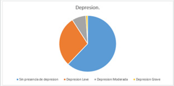
Figure 1: Patients with depression. Fuente. Zung & Conde Depression Scale. 62% of the patients did not present depression 34 patients, 16 patients had low depression 29%, also 8% of the patients had moderate depression which represented 4 patients also 2 of the patients representing 1% presented what could be considered as severe depression.
Lupinepublishers-openaccess-neurology-brain-disorders-journal

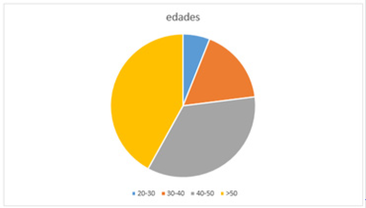
Figure 2: Most common ages seen during study. 42% of the patients presented depression were over 50 years old. 35% of the patients presented depression between 40-50 years of age. 17% of the patients that presented depression had between 30-40 years of age. 6% of the patients had between the ages of 20 to 30 years of age.
Lupinepublishers-openaccess-neurology-brain-disorders-journal

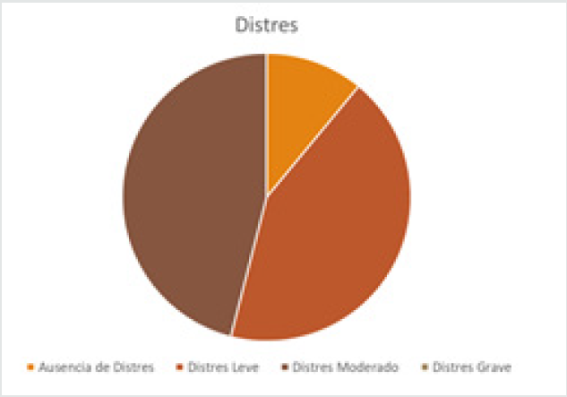
Figure 3: Patients that had some sort of stress evaluated by HADS scale.
a) 43% of the patients had lack of stress.
b) 46% of the patients had low level stress.
c) 11% of the patients had moderate stress.
d) 0% of the patients had severe stress
Lupinepublishers-openaccess-neurology-brain-disorders-journal

Justification

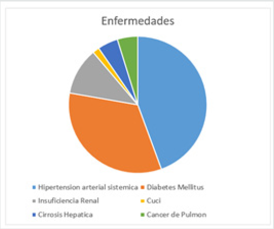
This study was conducted to observe what was the impact of being hospitalized and the relation it had with depression and anxiety in patient, we pretended to find viable date that allowed us to expose the hospital environment and the presence of disease, anxiety and depression (Figure 4) [11,12].

Figure 4: Most common diseases (12 patients didn’t have an illness):
a) Hypertension: 16
b) Diabetes Mellitus:12
c) Renal Insufficiency: 4
d) Ulcerative Colitis:1
e) Hepatic Cirrosis: 3
f) Lung Cancer: 3
g) Cervical Cancer:1
h) Fractures: 4
Lupinepublishers-openaccess-neurology-brain-disorders-journal

Results

Figure 1 Patients with depression. Fuente. Zung & Conde Depression Scale. 62% of the patients did not present depression 34 patients, 16 patients had low depression 29%, also 8% of the patients had moderate depression which represented 4 patients also 2 of the patients representing 1% presented what could be considered as severe depression (Figure 5,6).

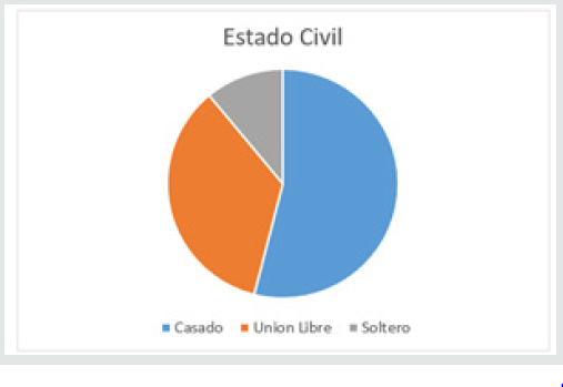
Figure 5: Civil Status:
a) Married: 54%
b) Separated: 35%
c) Single: 11%
Lupinepublishers-openaccess-neurology-brain-disorders-journal

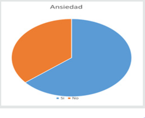
Figure 6: Patients that presented anxiety
a) 64% presented anxiety 35 patients
b) 36% did not present anxiety 21 patients
c) 86% No
d) 14% Yes
Lupinepublishers-openaccess-neurology-brain-disorders-journal

Conclusion

Referred to this subject we understand this two disorders are preventable and the patient if its treated and seeks help with time he can change his daily habits, our suggestions are that a stable lifestyle with a well-balanced diet consisting of fruit, vegetables, meat, daily exercise, stable relationships with family and friends, alongside no work stress, in addition to a good mental stability can lead to a good life and to prevent this type of disorders also to prevent chronic illness which were seen during this study on most patients that indicated feeling anxious or depressed, if they are in this state also to take medication on time and with the help of family members and friend.
a) We observe most patients presented a low depression level
b) Most patients presented anxiety
c) Most patients had a chronic illness
d) Patients over 50 years old presented higher depression levels
e) Most patients were married
Most patients had low level stress level.

Read More about this article: https://lupinepublishers.com/neurology-brain-disorders-journal/fulltext/depression-and-anxiety-frequency-in-patients-hospitalized-on-the-guadalajara-regional-military-hospital-in-the-month-of-april-2019.ID.000171.php

Read more Lupine Publishers Google Scholar Articles: https://scholar.google.com/citations?view_op=view_citation&hl=en&user=X9lN_1AAAAAJ&citation_for_view=X9lN_1AAAAAJ:PcT55Ow6fAIC


Monday, July 18, 2022

Swiss Cheese Pattern a Harbinger of Dementia or an Incidental Finding in an Unusual Case?


Abstract

Dilated Virchow Robin (VR) spaces are pial- line fluid filled structures which surround the walls of small penetrating vessels. In a severe form they develop a swiss cheese pattern or a cribriform pattern in straitum which may predispose to cognitive impairment. We report a patient with change in personality associated with diffuse atrophy, hypometabolism, microbleeds and swiss cheese striatum which is rare.

Read More about this article: https://lupinepublishers.com/neurology-brain-disorders-journal/fulltext/swiss-cheese-pattern-a-harbinger-of-dementia-or-an-incidental-finding-in-an-unusual-case.ID.000170.php

Read more Lupine Publishers Google Scholar Articles: https://scholar.google.com/citations?view_op=view_citation&hl=en&user=X9lN_1AAAAAJ&citation_for_view=X9lN_1AAAAAJ:FKYJxdYMdFIC

Friday, February 25, 2022

Lupine Publishers| Schizophrenia, Carbonyl Stress and Carnosine

 Lupine Publishers| Journal of Neurology and Brain Disorders


Abstract

Recent research suggests that schizophrenia is associated with the development of an advanced aging phenotype (carbonyl stress) and erythrocytes from schizophrenics also exhibit symptoms of cellular aging (increased levels of glycated proteins and ubiquitinated proteins), possibly due to excessive glycolysis-induced methylglyoxal (MG) generation. The endogenous dipeptide carnosine (beta-alanyl-L-histidine), which can delay cellular aging, suppress glycolysis and inhibit MG-induced protein glycation, also exerts some beneficial effects towards schizophrenia. Carnosine is present in human erythrocytes and the olfactory bulb (olfactory dysfunction is associated with schizophrenia). It is suggested that enhanced erythrocyte and olfactory carnosine levels may be more therapeutic towards schizophrenia, if carnosine was also administered intra-nasally to avoid serum carnosinase activity.


Keywords:Carnosine; glycation; methylglyoxal; erythrocyte; aging; nasal administration

Introduction


Schizophrenia and carbonyl stress

Many studies have indicated a relationship between schizophrenia and dysfunctional energy metabolism [1-3] whilst others indicate that carbonyl stress and generation of advanced glycation end-products (AGEs) accompany schizophrenia [4,5]. Furthermore, a recent study suggests that changes in glycolysis and accelerated cellular aging in glial cells contribute to the condition [6]. The glycolytic intermediates glyceraldehyde-3-phosphate and dihydroxyacetone-phosphate are the most likely sources of AGE formation due to their ability to spontaneously decompose into methylglyoxal (MG). MG is well recognized as a major glycating agent and is thought to be responsible for much macromolecular modifications associated with type-2 diabetes and age-related neurodegenerative conditions [7,8]. However, there is no clear evidence whether suppression of MG generation, via decreased glycolytic activity, has any effect on schizophrenia. The suggestion that schizophrenia seems to be associated with accelerated cellular aging [6] is supported by another recent observation reporting that erythrocytes obtained from schizophrenics contain elevated mounts of ubiquitinated proteins [9]. This might arise from either increased generation of targets for ubiquitination (e.g. aberrant polypeptides or denatured misfolded proteins), or decreased de-ubiquitinating activity, or decreased proteasomal proteolytic activity which would normally complete polypeptide destruction. Interestingly, MG and other agents responsible for carbonyl stress, also induce protein cross-linking which not only renders the target protein less susceptible to proteolytic attack but can also result in inhibition of proteasome activity generally [10]. Thus, it is conceivable that excessive glycolysis can provoke an aging phenotype (AGE accumulation and proteostatic dysfunction) via increased MG generation; such a relationship has been demonstrated in mice fed a high glycemic- index diet [11]. Never-the-less it is necessary to show whether glycation compromises proteostatic in erythrocytes from schizophrenics.


Erythrocytes and schizophrenia

A number of recent papers have revealed that erythrocytes obtained from patients with neurological problems, such as Alzheimer’s Disease (AD) and Parkinson’s Disease (PD), exhibit symptoms typical of aging cells in general. For example, compromised proteolytic activity and MG detoxification were detected in AD erythrocytes [12] and accumulation of aggregated protein occurs in red cells from PD patients [13]. Furthermore, dysfunctional energy metabolism, especially in relation to glycolysis culminating in carbonyl stress, are now regarded as characteristics of both AD and PD [14,15]. Therefore, it is not surprising that evidence of carbonyl stress is also accompanied by enhanced protein glycation [16] and accumulation of ubiquitinated proteins [9] in erythrocytes (and possibly other cells) obtained from schizophrenic individuals [17]. Moreover, one of the glycated proteins from “schizophrenic” red cells has been identified as a selenium-binding protein (SBP1) [18]; dysfunctional selenium metabolism has long been regarded as an important contributor to schizophrenia [19,20]. Selenium plays an important role in Sulphur metabolism required for synthesis of antioxidant enzymes such as glutathione peroxidase [21]. Thus, one is beginning to understand the relationship between AGE generation, carbonyl and oxidative stress and the apparently disparate biochemical attributes to schizophrenia.


Carnosine, carbonyl stress and schizophrenia

That erythrocytes can contain elevated amounts of MG and glycated proteins suggests the possibility that such red cells could become systemic sources of MG and AGEs to the brain and other tissues, following MG-induced eryptosis [22]. Consequently, it is important to consider whether suppression of carbonyl stress, not only in erythrocytes but in astrocytes and glia, could possibly be a therapeutic strategy. The naturally occurring dipeptide carnosine (beta-alanyl-L-histidine) has been shown to suppress glycolysis in cultured cells [23,24], delay replicative senescence [25], stimulate proteolysis of long-lived proteins in late passage cells [26] and inhibit AGE formation [27]. Furthermore, there is one study showing that schizophrenics subjected to dietary supplementation with carnosine exhibited some beneficial effects [28], possibly due to the dipeptide’s pluripotent properties [29]. It is also interesting to note that

a) Olfactory dysfunction is also associated with schizophrenia [30,31] and

b) Carnosine is enriched in the olfactory bulb [32].

Thus, one has to consider whether raising olfactory carnosine levels could also be useful. However, all studies employing dietary carnosine supplementation are subject to the problem of the presence of serum carnosinase activity which would destroy the dipeptide [33]. There is an alternative route however, which is to use an intra-nasal approach. This could involve a nasal spray of a carnosine solution; another approach could involve use of carnosine powder. Indeed “snorting” carnosine could be far more useful than most white powders some people use, be it illegal drugs or “medicinal snuff “of old. In fact, intra-nasal delivery of potential therapeutic agents is currently being explored [34] with respect to neurodegenerative conditions, as proposed many years ago [35].

Carnosine has been detected in human erythrocytes [36] but in lower amounts when obtained from elderly individuals [36]. It is presumed that red cell carnosine is synthesized (from betaalanine and histidine) during erythropoiesis. Consequently, it would be useful to determine whether dietary supplementation with carnosine or beta-alanine raises erythrocyte carnosine levels and whether there are any beneficial effects with respect to the recognized changes in “schizophrenic” erythrocytes. Additionally, it is suggested that any carnosine (dietary or nasally administered) supplementation period should last for at least 120 days to ensure maximal numbers of carnosine-enriched erythrocytes. It has been proposed that excessive and continuous glycolysis in erythrocytes enhances red cell MG levels, and thus also facilitate delivery of erythrocyte MG to the tissues including the brain [22]. Consequently, it will be also important to determine whether such supplementation protocols decrease carbonyl stress and MG levels not only in red cells but the tissues generally including glia [6].

  Read More Lupine Publishers Journal of Neurology and Brain Disorders: https://brain-disorders-lupine-publishers.blogspot.com/

Friday, February 4, 2022

Lupine Publishers| Primary Lateral Sclerosis, Report of a Case and Bibliography Revision

 Lupine Publishers| Journal of Neurology and Brain Disorders

Abstract

Introduction: Primary lateral sclerosis is a rare disease involving the upper motor neuron, producing a bulbospinal spasticity. The course of the disease is insidious and progressive, usually starting with the lower extremities, and later becoming a tetrapyremidal syndrome. As a rare disease, diagnosis in most cases is exclusionary, and the patient should be studied extensively, clinically, including a thorough medical history, laboratorial and with the relevant cabinet studies.

Classic Case: It is male patient who starts his clinical picture about a year ago with weakness in left pelvic limb, subsequently accompanied by pain and paresthesias, manifesting the same symptomatology later in the contralateral leg and upper left limb. Currently is added index and middle toe hypoesthesia, moderate tremor in left arm, with the contrast of the middle toe over the ring of said hand. It has an inability to lift light objects for short periods of time, as well as fatigue in short periods of time when performing daily activities, which greatly limits their daily life.

Conclusion: Motor neurone diseases are divided into two groups, and in the case studied, the upper motor neuron is exclusively affected. As it is a rare disease, with a low incidence, multiple differential diagnoses will be considered before concluding in it, considering it a diagnosis of exclusion. The natural history of the disease will always have a grim outcome, with poor prognosis for life and function, despite the measures taken to change the course of the disease.

Keywords:Motoneuron; upper; spastic; resonance

Introduction

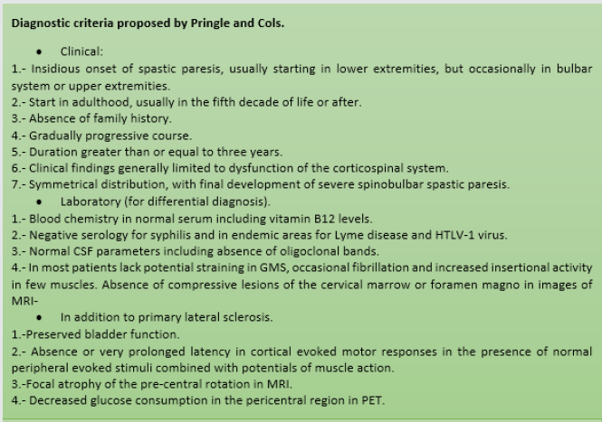
Primary lateral sclerosis is a rare disease involving the upper motoneuron, which is characterized by a progressive bulbospinal spasticity, with selective degeneration of pyramidal neurons located in the precentral convolution [1]. Primary lateral sclerosis makes up approximately 1% to 4% of all patients with motor neuron diseases [2]. The onset of the disease is insidious, with a slow and progressive spastic paralysis, usually starting at the lower extremities, and then becoming a tetrapyremidal syndrome. On average, patients are estimated to have a life prognosis greater than 10 years from the onset of symptoms. Hyperreflexia, moderate weakness, dizziness, lack of coordination may also occur within the clinical picture. In the physical examination you can find spasticity, increased osteotendinous reflexes, and as a predominant sign, stiffness [2,3]. Diagnosis is usually performed clinically, relying on MAGNETIC resonance imaging, among other studies, to rule out other pathological entities, showing in this study a marked atrophy in the primary motor cortex [3]. In some studies, diffusion magnetic resonance imaging has been used, showing a functional increase in cerebral axonal activity, especially the brain-cerebellar, which could explain an adaptive process through functional neuroplasticity, however, the prognosis of the disease remains unfavorable and irreversible despite these changes [4]. Although there are some diagnostic criteria for the disease, as it is a rare entity, not all inclusions for it are always met, so it is usually a diagnosis of exclusion [5,6] (Figure 1).

Figure 1: Diagnostic criteria proposed by Pringle and Cols.

Lupinepublishers-openaccess-neurology-brain-disorders-journal

Clinical Case

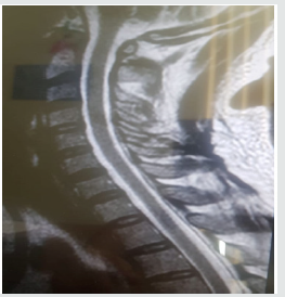
They are a 42-year-old male patient, who started his clinical picture about a year ago, when when he is parading, I notice some weakness in left pelvic limb, progressing over the course of the days to diffuse pain and local paresthesias, and subsequently having the same problem in the contralateral leg and then to upper left limb throughout, progressively to date. Currently, adding to the above, it has hypoesthesia of index and middle toe, stiffness in all the arcs of the movement of the affected limbs, moderate tremor in the left arm, with the oversetting of the middle toe over the ring of said hand. Symptomatology is exacerbated in the mornings. It has an inability to lift light objects for short periods of time, as well as fatigue in short periods of time when performing daily activities, which greatly limits their daily life. It denies important here family backgrounds, as well as surgeries, drug addictions, or known diseases (Figure 2).

Figure 2: Cervical spinal MRI where you see a normal morphology without compromise spinal cord.

Lupinepublishers-openaccess-neurology-brain-disorders-journal

Discussion

Primary lateral sclerosis is a rare disease affecting the upper motor motoneuron of the primary motor area (pre-central circumvolution),characterized by having an insidious onset in people with no known risk factors, with a clinical picture in which a spastic paresis of lower limbs of onset predominates that can be generalized until tetra pares progressively increasing over the years and which is usually a long-evolving disease. Diseases of the motor neuron are divided into those that affect the upper and lower, in this case we focus on the first case, which are located inside the cerebral cortex and send axons that form the pyramidal pathway, to later defuse and finally connect to the spinal cord. The manifestations secondary to the lesion of the upper motoneuron are as follows: spastic paralysis, amyotrophy (by disuse), absence of tracing, exalted myotatic reflexes and extensive plantar response.

As it is a disease with a low incidence and few reported cases, it should never be considered as a diagnosis of first instance, having to rule out other differential diagnoses, so over time various diagnostic criteria have been defined to realize the disease, however, as well mentioned, it is a rare disease, so no definitive consensus has been created for the diagnosis and scrutiny of the disease, however, we can lead our diagnostic approach means of any of these, such as the criteria of pringle and cabbage, through a well-established clinic, conducting a thorough clinical history, laboratory studies, ranging from general analyses such as hematic biometry, blood chemistry, functional tests, to cytological examinations of cerebrospinal fluid, quantification of vitamin B12 in serum, trepamic tests, as well as cabinet studies such as electromyography and MRI, where the absence of other pathological entities is verified and in the latter study a marked cortical atrophy of the pre-central turn. Despite the above, it should be noted that the disease may not follow a specific pattern within its natural history, so it may vary the presentation and sequence of them.

Conclusion

Motor neuron diseases are divided into two groups, and in the case studied, the upper motoneuron is exclusively affected, differing from each other by the form of presentation and clinical manifestations. As it is a rare disease, with a low incidence, multiple differential diagnoses will be considered before concluding in it, considering a diagnosis of exclusion, and thus, a thorough medical history should be made with a proper examination conducting laboratory and cabinet studies relevant to this situation. Although it is a long-term disease with a longer life expectancy than amyotrophic lateral sclerosis, the natural history of the disease will always have a grim outcome, with poor prognosis for life and function, taking into account there is no cure for the disease other than supportive treatment.

 Read More Lupine Publishers Journal of Neurology and Brain Disorders: https://brain-disorders-lupine-publishers.blogspot.com/


Lupine Publishers| Depression and Anxiety Frequency in Patients Hospitalized on the Guadalajara Regional Military Hospital in the Month of April 2019

  Lupine Publishers| Journal of Neurology and Brain Disorders   Abstract Observe and Identify patients that presented depression ...