Journal of Neurology & Neurosurgery-Lupine Publishers
Abstract
Keywords: Cerebrocerebellar Circuit; Hypothalamus; Cerebellum; Multilayered Fibers; Histamine
Introduction
The HCC consists of a descending limb, formed by the hypothalamocerebellar fibers, and an ascending limb, formed by cerebellohypothalamic fibers [3]. The hypothalamocerebellar fibers arise from widespread nuclei/regions of the hypothalamus including the paraventricular nucleus [4,5], the ventromedial nucleus [4,6], the tuberomammillary nucleus and the lateral region of the posterior zone [4,7]. The hypothalamocerebellar fibers run along the midbrain tegmentum, and reach the cerebellum, mainly through the ipsilateral superior cerebellar peduncle; in the cerebellum, they send collaterals to all the cerebellar nuclei, and terminate in the cortex of all the cerebellar lobes [8,9]. On the other hand, the cerebellohypothalamic fibers originate from all the cerebellar nuclei, particularly from the fastigium nucleus, enter the superior cerebellar peduncle, and ascend in the midbrain tegmentum up to the hypothalamus, where they terminate on the same nuclei and regions at the origin of the descending limb [10].
Studies have indicated that the hypothalamocerebellar fibers end in the cerebellar cortex with different ways from the other fibers afferent to it: precisely, they end on all the cortical layers, multilayered fibers, and synapse on granule neurons, Purkinje neurons and interneurons of the molecular layer [9,10]. The behavior of these fibers is clearly different from that of the mossy or climbing fibers, which, instead, selectively end in the granular layer or in the molecular layer [11]. Another difference between the multilayered fibers and the mossy and climbing fibers is that some multilayered fibers use histamine as a chemical neurotransmitter, while the mossy and climbing fibers predominantly use glutamate[12]. Accordingly, physiological studies have reported that histamine exerts postsynaptic excitation on granule and Purkinje neurons of the rat cerebellar cortex [13,14], and a study conducted by PET techniques has revealed histamine receptors in the human cerebellar cortex [15].
It is noteworthy that the tuberomammillary nucleus, the only proven source of histaminergic fibers in the central nervous system, is also one of the hypothalamic nuclei at the origin of hypothalamocerebellar fibers [16]. Aim of the present study was to analyze the fine distribution of histamine-containing axon terminals in the human cerebellar cortex with the goal of defining the morphological organization of its histaminergic nervous circuits and evaluating the interactions between the histaminergic terminals and the wide distributed GABAergic and glutamatergic terminals. The study was carried out by light microscopy immunohistochemical techniques based on a highly specific polyclonal antibody against histamine.
Material and Methods
The fragments were fixed by immersion for 2-4 h depending on their size in saturated solution of picric acid containing 4% paraformaldehyde [17] and embedded in a semi-synthetic paraffin wax. Paraffin blocks were serially cut into 5-mm sections orthogonally oriented towards the cerebellar surface. The sections were incubated with polyclonal antibodies, developed in rabbit using histamine conjugated to succinylated KLH as immunogen (Sigma, MO, USA), diluted 1:50 in PBS. The immunoreactions were revealed by the streptavidinperoxidase complex (Dako LSAB kit, Dako, CA, USA), using 3-amino-9-ethyl-carbazole (Vector Laboratories, CA, USA) as a chromogen.
Negative controls: total abolition of the immunolabeling was obtained by treating sections with non-immune serum or omitting the primary antibodies.
Results
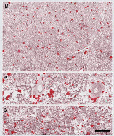
In the molecular layer, only a small number of isolated immunoreactive puncta was observed in the neuropil or on the surface of neuronal bodies and processes of the layer; they appeared uniformly concentrated throughout the layer. In the Purkinje neuron layer, some puncta were observed on the body surface of a few of Purkinje neurons, concentrating on their deep pole (preaxon region). In the granular layer, a relatively larger number of immunoreactive puncta was observed, isolated or gathered in small groups, in the islet of neuropil comprised among the granules (Figure 1).
Figure 1: Histamine immunoreactivity was detected in
puncta distributed in all the layers of the human cerebellar
cortex (tonsilla). In the molecular layer (M), a small number
of isolated immunoreactive puncta was observed mainly
in the neuropil. In the Purkinje neuron layer (P), some
puncta were observed on the body surface of a number of
Purkinje neurons, concentrating on their deep pole. In the
granular layer (G), the puncta were observed isolated or
gathered in small groups in the islet of neuropil comprised
among the granules. Scale bar: 40x.


Discussion
In other terms, the histamine-immunoreactive terminals act on selected neuronal groups intervening in the processing of afferents from hypothalamus and originating efferents constituting the ascending limb of the HCC. The latter is firstly represented by corticonuclear fibers projecting on cerebellar nuclei. Since they originate from all cerebellar lobes, these corticonuclear fibers project on all the cerebellar nuclei, namely the fastigium, interpositum and dentate nucleus. These data agree with those indicating an involvement of all the cerebellar nuclei in the HCC [8]. The selected nuclear neurons give rise to the cerebellohypothalamic fibers, the closing section of the ascending limb. It is interesting that neurons in all the cerebellar nuclei received histaminergic terminals from collaterals of the hypothalamocerebellar fibers [18-20]. In practice, histamine would be important both in regulating some intrinsic circuits of the cortex, from which activity the efferents destined to the nuclei arise, and in regulating the activity of some nuclear neurons, at the origin of efferents to the hypothalamus.
The descending limb of HCC and NCC, the two components of the cerebrocerebellar circuit, terminates with different distributional patterns in the cerebellar cortex: HCC terminates ubiquitously, with few histaminergic terminals belonging to multilayered fibers, NCC terminates in the cortex of the cerebrocerebellum, with a greater number of glutamatergic terminals belonging to mossy fibers. It is likely that interactions between histaminergic terminals (i.e., HCC) and glutamatergic terminals (i.e., NCC) occur in the regions of cerebellar cortex where they converge. Such interactions would underlie the combined role of the cerebellum in the regulation of somatic and non-somatic functions. These results open new perspectives in the interpretation of the physiopathology of neurological and psychiatric disorders depending on dysfunction of the cerebellum and offer new therapeutic possibilities. In this view, the use of histamine has already been proposed for the treatment of cerebellar ataxia [21,22]. Further applications are needed in light of the involvement of the cerebellum in an increasing number of pathologies, including disorders of motor, psychic and visceral functions.
For more Lupine Publishers Open Access Journals Please visit our website:
https://lupinepublishers.us/
For more Open Access Journal on Online Journal of Neurology and Neurosurgery articles Please Click Here:
https://lupinepublishers.com/neurology-brain-disorders-journal
To Know More About Open Access Publishers Please Click on Lupine Publishers


No comments:
Post a Comment